|
|
Image Formation for an In-Vivo Ultrasonic MicroprobeBy Mark A. Haun. The ultrasonic microprobe project is working to develop tiny ultrasound transducers that can be fabricated on a needle and operated in-vivo at high frequencies, with the eventual goal of resolving individual cells. A variety of synthetic aperture and tomographic imaging techniques are being explored to reconstruct the three-dimensional tissue volume surrounding such a probe. Related work will investigate methods for the compensation of phase errors caused by tissue inhomogeneities, as required for coherent imaging. Other challenges caused by the nonlinear propagation of sound may also need to be addressed. One imaging concept uses a single focused transducer on the surface of a needle and builds on earlier work with synthetic aperture image formation with virtual sources. Given a set of pulse echoes recorded at various needle rotation angles and vertical positions, good resolution is achieved on the cylindrical surface defined by the transducer focal length and needle motion. Fourier-based synthetic aperture techniques can then be used to reconstruct the tissue reflectivity on more distant cylindrical shells with comparable resolution and reasonable computational effort.
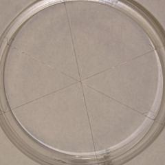
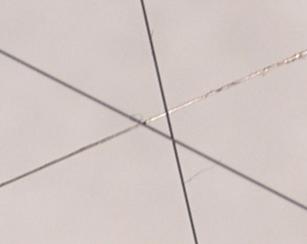
0.1-mm wire target (left) and center detail (right). The central area of this target was imaged using a 2.25-MHz, f/1.33 transducer scanning over a section of a cylindrical shell 15 mm high and 18 degrees wide. The center of the target was about 10 mm beyond the transducer focus.
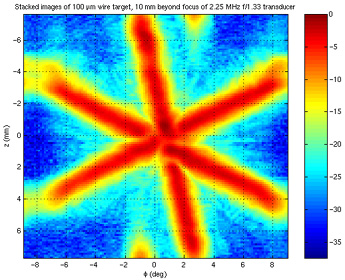
Left: Animated fly-through of the reconstructed 3-D volume from the above experiment. Each of the 50 frames is an unfolded cylindrical shell going 50 microns deeper. Thus, the total volume shown here is a cylindrical sector 15 mm high by 18 degrees wide by 2.5 mm deep, located about 50 mm from the cylinder axis. Right: A stacked, log-scaled version of the data. As predicted by theory, the resolution is on the order of one wavelength at 2.25 MHz. The slight wrap-arounds are a processing artifact.
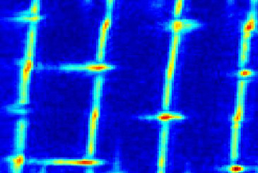
An ordinary piece of 1/16-inch-mesh aluminum screen was imaged 5 mm past the focus of a 15-MHz, f/1.5 transducer (left). The reconstructed image (right) clearly shows the mostly straight vertical wires. Due to the over/under weaving pattern of the horizontal wires, only the overcrossings are normal to the ultrasonic beam and display well. BRL Projects >> |
|||||||||||||||||||||||||||||||||||||||||||||||||||||||||||||||||||||
| Bioacoustics Research Lab. |